


It will be very important and exciting to see this new therapy being given to patients one day.
Brian W. Pogue, PhDInspired by a presentation on Cherenkov light use in basic nuclear medicine research, it was apparent to Brian W. Pogue, PhD, co-Director of the Translational Engineering in Cancer Research Program at Dartmouth Cancer Center, that there would be much more Cherenkov light coming from radiation therapy, and that this light could be harnessed to visualize the beam during patient treatment.
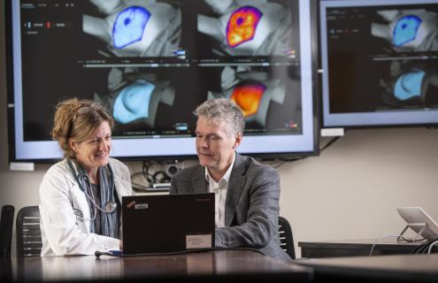
The Cherenkov effect occurs when radiation beams interact with tissue, such as skin, producing a small light emission from the surface. With a camera provided by a post doc, Pogue and Dartmouth Cancer Center colleague David Gladstone, ScD, chief of Clinical Physics in Radiation Oncology, suggested turning off the lights and photographing light emissions from phantoms. “This was the very start of it at Dartmouth,” says Pogue.
Basic beam imaging studies turned into veterinary studies, initiating the first human imaging study in 2014, publication of which spurred interest in developing better visualization for everyday radiotherapy.
Commercial pipeline milestones
The team’s first patent showing capability of imaging radiation dose non-invasively in water tanks and on tissue phantoms allowed for formation of DoseOptics, LLC, licensed by Dartmouth in a biotech startup partnership program.
Pilot funding from the National Cancer Institute (NCI) allowed for initial design of a new camera system and software by faculty Petr Bruza, Michael Jermyn and Venkat Krishnaswamy from Dartmouth’s Thayer School of Engineering. Key partnerships and subcontracts with Radiation Oncology at Dartmouth Hitchcock Medical Center (DHMC) for access to machines called linear accelerators (LINACs) that deliver radiation therapy, and LINAC users were critical in the years-long development process.
Today, DoseOptics has earned more than $8 million in NCI Small Business Innovation Research (SBIR) funding and introduced two FDA 510(k)-cleared products to the market:
The initial BeamSite, a single camera system received FDA 510(k) clearance in December 2020, coincidentally, the same day that the first COVID-19 vaccine was approved by the FDA. In November 2021, an enhanced multi-camera system called doseRT received clearance and was marketed commercially.
Over the past five years, DoseOptics has committed more than $1 million to Dartmouth in support of joint Cherenkov development projects. In that time, the team has evolved the camera system into an extremely stable, sensitive technology capable of imaging very low light signals from the most advanced radiotherapy treatment deliveries.
BeamSite systems installed in all Dartmouth Cancer Center patient radiation therapy rooms continue to provide clinical feedback to inform adjustments resulting in delivery of the best radiotherapy possible for patients, while also keeping the device simple and easy to use. Completion of the first pilot study of radiotherapy imaging with Cherenkov confirmed the value that this imaging brings to the clinic.
Cherenkov-activated nanomicelle chemotherapy (CHANT)
Eliciting the value of Cherenkov light did not end with imaging cameras. Cherenkov-activated nanomicelle chemotherapy (CHANT) can be used to encapsulate chemotherapy, making it extremely sensitive to ultraviolet light.
“Because Cherenkov light is largely ultraviolet, we could release chemotherapy locally in tumors by X-ray radiotherapy, and the Cherenkov light would activate the release,” explains Pogue. The team studied delivering chemotherapy in nanomicelle packages directly into tumors, and then releasing the chemotherapy by irradiating the tumors with a standard dose of radiation.
Findings from several related studies were published and the team is planning more extensive testing.
FLASH ultra-high dose rate radiotherapy
Other advancements in radiation include ultra-high dose rate (UHDR): extremely fast radiation delivery in short bursts. UHDR has been found to be effective in killing tumor tissue, but with up to 50 percent less damage to normal tissues. This has been called the ‘FLASH’ effect.
A large team of Dartmouth Cancer Center and Dartmouth faculty ramped up research and development in FLASH radiotherapy during the COVID-19 shutdown, using the time to virtually confer with leading academics from around the world.
FLASH work is also supported by Prouty funds. This year, Chun-Chieh Lin, MD, PhD, with mentors Pogue and radiobiologist P. Jack Hoopes, DVM, PhD, are diving into the basic epigenetic mechanisms of how normal and tumor cells react to FLASH in their Prouty Pilot project.
LINAC conversion development
Realizing the capacity to develop FLASH in existing radiotherapy centers was possible, the Medical Physics team led by Rongxiao Zhang, PhD, Gladstone and Bruza led the conversion of a standard LINAC to ultra high-dose capability. The FLASH LINAC conversion process has since been reduced from more than one hour to less than five minutes and tested more than 100 times in early-phase study mode offline.
Hoopes and his lab have also studied the intriguing FLASH phenomenon in mice skin and pig skin and in a Phase I safety trial in veterinary dogs with natural cancer whose owners consent to enrollment. The studies have shown promise that safe delivery can be achieved. The future of FLASH may therefore include human safety trials.
FLASH radiotherapy oxygen measurements
One core thought about why the FLASH effect occurs is oxygen depletion in tissue from the rapid dose delivery. Reinforcing why Dartmouth is a leader in measuring oxygen in vivo, the team directly measured the oxygen changes that occur during FLASH radiotherapy, and, through the first in vivo measurement of this phenomena ever documented, showed that these brief changes in oxygen could be observed.
FLASH human clinical trial planning
Holding the only method of visualizing and recording radiotherapy beam delivery to patients—a product of the collaborative synergy embedded in the core of the Dartmouth Cancer Center, Dartmouth and DHMC research ecosystem—the diverse clinical research team is well-staffed, well-experienced and well-supported to spend the next few years standing up the very first FLASH human clinical trial of advanced-stage skin lesions. The trial would serve to verify dose delivery to every single patient by directly imaging the delivery with Cherenkov cameras. “It will be very important and exciting to see this new therapy being given to patients one day,” says Pogue.