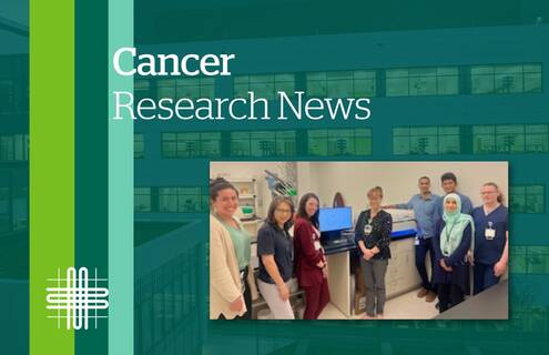
Chromosomes are the structures in our cells that carry our genetic information. They are like a blueprint for how our bodies are built. Certain structural abnormalities or physical differences in the chromosomes, such as the order in which the genes are in, can lead to genetic disorders or diseases such as cancer. For the last 40 years, standard-of-care approaches for diagnosing blood cancers due to chromosome variation have been labor-intensive and limited in resolution.
Dartmouth Health is one of the first academic medical centers in the country, and the first in New England, now taking standard of care to the next level. Launched in May, Optical Genome Mapping is first-of-its-kind digital karyotyping technology to improve the diagnosis of hematologic cancers, including leukemias and lymphomas.
Validated by the Cytogenetics Laboratory, under the Clinical Genetics and Advanced Technology (CGAT) section of the Department of Pathology and Laboratory Medicine, Optical Genome Mapping is taking extremely close-up images, or digital karyotypes, of chromosomes and then analyzing the images with specialty computer software to detect abnormalities across the entire map of human chromosomes.
“This test can produce comprehensive chromosome structure and copy number variation detection at 1,000-fold resolution compared to current approaches - a resolution that was previously not possible,” says Wahab A. Khan, PhD, FACMG, Director of Cytogenetics and Molecular Genetics and Associate Director of CGAT. “This has allowed us to now clarify the well-known blind spots that are part of standard-of-care testing, as well recognize important gene content involved in hematological malignancies on a genomic scale.”
The digital karyotype process by Optical Genome Mapping incorporates fluorescent labels into very long strands of DNA. These labels are then computationally mapped to the reference genome. Any areas where genomic information has been shuffled, lost, or gained are flagged, offering clues as to how and why a particular cancer has developed.
“For example, in chronic myelogenous leukemia, we know that there's a very specific type of chromosome abnormality that takes part of one gene and shuffles it next to another gene (called a ‘translocation’ or ‘fusion event’). But those two genes should not be next to each other and that causes overproduction of abnormal cells,” explains Khan. “This test can find abnormalities like this on an entire genome-wide level – a vast landscape of abnormalities with diagnostic and prognostic implications that would have gone unrecognized by more conventional methods.”
Having rigorously validated the platform for use in chronic and acute adult blood cancers, the Clinical Cytogenetics lab under CGAT has worked with providers in Hematopathology to get the test into the clinic and incorporated into patient electronic medical records. Although still early, the benefit for patients is that the test can categorize risk and prognosis much better, provide more clarity in diagnostic chromosome findings, and allow the potential for more informed treatment plan decisions.
The Cytogenetics team in CGAT aims to optimize and validate the protocol for additional cancers, such as fresh-tissue lymphomas, as well as other indications and cell types such as building digital karyotypes for multiple miscarriages and infertility.
***
Wahab A. Khan, PhD, the Director of Cytogenetics and Molecular Genetics and Associate Director of Clinical Genetics and Advanced Technology at Dartmouth Health, and Assistant Professor of Pathology and Laboratory Medicine at the Geisel School of Medicine at Dartmouth. His research interests include cytogenomics, molecular diagnostics, chromosome biology and medical genetics and genomics.