


ZEISS LSM 800 laser point scanning confocal microscope with Airyscan enhanced resolution
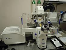
Four Diode lasers for confocal imaging:
- Diode laser (405 nm, 5 mW); class 3B (DAPI, Hoechst, PPIX, Filipin, paGFP)
- Diode laser (488 nm, 10 mW); class 3B (GFP, CFP, FITC, AF488, YFP)
- Diode (SHG) laser (561 nm, 10 mW); class 3B (Cy3, TxRed, AF568, AF594)
- Diode laser (640 nm, 5 mW); laser class 3B (AF647, CY5, APC)
Confocal detectors:
- Two Gallium Arsenide Phosphide (GaAsP) PMTs
- One additional GaAsP Airyscan enhanced resolution PMT detector for 63× objective
Lumencor LED Spectra X Light Engine- for wide-field fluorescence microscopy:
- Excitation 395/25
- Excitation 470/24
- Excitation 550/15
- Excitation 640/30
- Excitation 720/20
Wide-Field Bright-field and Fluorescence Detector:
- Hamamatsu C11440-22CU Digital CMOS camera ORCA-Flash4.0
Electronically switchable illumination and detection module (ESID) for transmitted-light imaging.
Zeiss AxioObserver wide-field inverted fluorescence microscope
Objectives available:
- 10X Plan-Apo/0.45 NA
- 20X Plan-Apo/0.8 NA with DIC capability
- 40X Plan-Apo/1.3 NA Oil with DIC capability
- 40X C-Apo/1.2 NA Water Immersion
- 63X Plan-Apo/1.4 NA Oil with DIC capability
Live Cell Incubation System:
- Full plexiglass enclosure, heating stage, and enclosure heater for 37ºC live imaging
- CO2 control system
- Definite Focus 2 autofocus
Akoya Vectra® 3 automated quantitative pathology imaging system
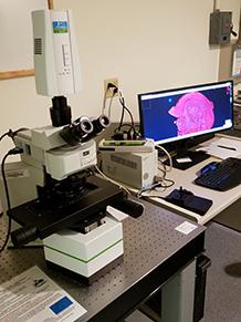
This slide scanner and associated software for either immunofluorescence (IF) or immunohistochemistry (IHC) slides is located in Borwell 338 West. The Olympus BX51 upright fluorescence microscope is equipped with an automated 6-slide stage. The imaging system allows imaging and measurement of weakly expressing and overlapping biomarkers within a single H&E, IHC or IF intact thin tissue sections or tissue microarrays. Vectra, Phenochart, and inForm software allow control of multiplexed biomarker imaging and quantitative analysis.
Tissue thin sections or TMAs can be labeled with IF or IHC stains or with conventional stains such as H&E and trichrome. When using IF or IHC stains, multiple proteins can be measured on a per tissue, per cell, or per cell compartment (e.g., nuclear, cytoplasmic) basis. Multi-spectral imaging using the Nuance camera allows unmixing of highly overlapping spectral signals and correction for autofluorescence.
Phenochart software permits contextual viewing and annotation capability of whole slide scans created using the 4X or 10X objectives. Composite images are automatically generated from the series of tiled images covering the slide coverslip area. Users can navigate around the stored digital image and identify areas of interest for further higher-resolution multispectral image acquisition using 20X or 40X objectives.
Objectives:
- 4X UPlan SApo NA 0.16 WD 13.0 mm
- 10X UPlan SApo NA 0.40 WD 3.1 mm
- 20X UPlan SApo NA 0.75 WD 0.6 mm
- 40X UPlan SApo NA 0.95 WD 0.18 mm
Agilent Cytation 5 real-time, quantitative live-cell analysis system with BioSpa8 incubator, gas mixture control and automated drug delivery system
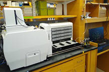
The Cytation 5 is an automated microscope/camera system that allows live cell imaging over extended periods of time. Long-term time course experiments can be performed using up to eight multi-well plates or chambered slides, shuttled between the long-term BioSpa8 incubation unit and the Cuytation 5 imaging unit by an automated robot arm. A well plate scratch module is available for monolayer scratch healing assays. Incubation controls for temperature, CO2 and nitrogen displacement to achieve hypoxia are available in both the imaging unit and the long-term incubation unit. Quantification of cell proliferation, chemotaxis, or apoptosis is possible using integrated Gen5 system software.
Objectives:
- 4x high contrast/fluorescence
- 4x phase/fluorescence
- 10x phase/fluorescence
- 20x phase/fluorescence
- 40x phase/fluorescence
Exported image formats:
- JPEG
- PNG
- TIFF
- RAW
Excitation/emission cubes:
| Name | LED | Ex (center/width) | Dichroic | Em(center/width) |
|---|---|---|---|---|
| DAPI | 365 nm | 377/50 nm | 409 nm | 447/60 nm |
| GFP | 465 nm | 569/35 nm | 497 nm | 525/39 nm |
| RFP/PI | 523 nm | 531/40 nm | 568 nm | 593/40 nm |
| TxRed | 590 nm | 586/15 nm | 605 nm | 647/57 nm |
| Cy5 | 623 nm | 628/40 nm | 660 nm | 685/40 nm |
| Cy5.5 | 655 nm | 647/57 nm | 695 nm | 794/160 nm |
| Cy7 | 740 nm | 716/40 nm | 757 nm | 809/81 nm |
Olympus IX-83 with PRIMO photopatterning module
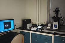
The PRIMO is a micropatterning / microfabrication bioengineering system consisting of a UV photo ablation/curing device capable of projecting an arbitrary pattern with up to 256 grey levels of intensity controllable by user drawn .PDF or .PNG file inputs down to micron resolution. Using Alveole Lab reagents, users can convert these designs into cell culture substrates in flat or 3D polymerized hydrogel formats.
- PRIMO photopatterning device
- Zero Drift focus module
- ASI MS-2000 with XY-Axis Heidenhain Linear Encoders
- Prior Lumen 200 widefield fluorescence light source
- Andor Sona scMOS camera
- Dell Precision T5600 Computer and Leonardo photopatterning software
Olympus IX-73 inverted fluorescence microscope with Olympus DP73 computer- controlled CCD camera
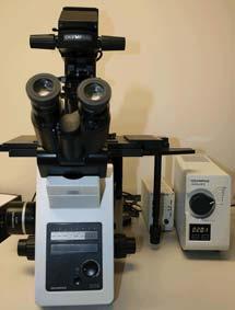
This inverted fluorescence microscope system allows imaging of multi-well plastic culture plates using long working distance 20X and 40X objectives. High-quality imaging of coverslip covered samples can be done using the 20X and 40X dry objectives or with oil immersion 40X and 100X high NA PlanApo objectives. The microscope has a wide array of fluorescence filter cubes in the eight-position turret to allow the use of fluorophores such as DAPI, GFP, TxRed, mCherry, Cy5, and IR Dye 800..
Objectives available:
- UPLFLN4X; U PLAN FLUORITE 4X OBJECTIVE, NA 0.13, WD 17MM
- UPLSAPO10X2; U PLANS S-APO 10X OBJECTIVE, NA 0.4, WD 3.1MM
- LUCPLFLN20XPH; LWD U PLAN FL PHI OBJ, NA 0.45, WD 6.4-7.6MM,W/CC
- LUCPLFLN40XPH; LWD U PLAN FL PH 2 OBJ, NA 0.6, WD 2.7-4.0, W/C-COL
- UPLFLN40XO; U PLAN FLUORITE 40X OIL OBJ, NA 1.30, WD 0.2MM
- UPLSAPO100XO; U PLAN S-APO 100X OIL OBJ, NA 1.40, WD 0.12
Fluorescence filter cubes available (8 positions):
- DAPI-5060C-OFF-ZERO; EX377/50, EM447/60, DM409
- TXRED-4040C-OFF-ZERO; EX562/40, EM624/40, DM593
- GFP-3035D-OFF-ZERO; EX472/30, EM520/35, DM495
- ET Cy5 longpass, EX620/60, EM660lp, DM660lpxrxt
- ET LI-Cor for IR Dye 800, EX740/40, EM780lp, DM 770lpxr
- ET DAPI/FITC/Texas Red
- ET EGFP/mCherry
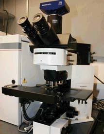
Olympus BX 50 upright fluorescence microscope
Objectives available:
- UPlanFL 10X/0.3
- UPlanFL 20X/0.5
- UPlanFL 40X/0.75
- Zeiss Achroplan 100X/1,25 oil can be placed into the turret on request
Fluorescence filter cubes:
- DAPI:ex-BP360-370, DM400,em-BA420
- FITC:ex-460-490, DM505, em-BA515IF
- Rhodamine:ex-BP520-550, DM565, em-BA580IF
Zeiss M2Bio stereomicroscope
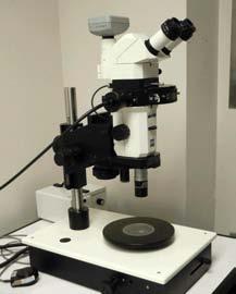
This stereomicroscope system provides low magnification long working distance (stereo) imaging with an 11X zoom. Field widths as large as 2.5 cm can be imaged. The microscope converts to monocular imaging with 10X or 20X LWD objectives and the 11X zoom. Trans or reflected Bright field and fluorescence imaging is possible. Images are recorded with either a B/W or color digital camera using Zeiss Axiovision software.
Objectives available:
- 0.6X
- 1.0X
- 1.6X
- 10X 0.45 NA f=200
- 20X 0.6 NA f=200
Fluorescence cubes available:
- Q dot 705 ex 460SPUVv2; bs 475DCXRU; em D705/20
- Coumarin ex D405/10, bs 425DCLP; em D460/50
- cGFP ex D436/20; bs 455DCLP; em D480/40
- GFP ex HQ470/40; bs Q495LP; em HQ525/50
- yGFP ex HQ500/20; bs Q515LP; em HQ535/30
- TRITC ex D540/25; bs 565DCLP; em D605/55
- Cy5 ex HQ620/60; bs Q660LP; em HQ700/75