


Location and access
Borwell 338 West
One Medical Center Drive
Lebanon, NH 03756
(603) 646-5503
Hours: Monday to Friday, 9 am to 5 pm
Authorized users have 24/7 access.
Overview
The Dartmouth Cancer Center Microscopy Shared Resource (DCC MSR) provides access to advanced imaging instrumentation and image analysis software to Dartmouth Cancer Center members, and all members of Dartmouth College and Dartmouth Hitchcock Medical Center communities.
The instrumentation available include point scanning confocal microscopy and Airyscan super-resolution, automated multi-spectral image acquisition and spectral unmixing of high-plex labeled slides, high throughput phase contrast and widefield fluorescence imaging of live cells, conventional widefield fluorescence and transmitted light imaging, and a UV light-based micropatterning and microfabrication bioengineering system. Workstations for image analysis are available within the facility.
To enable effective and safe use of the MSR resources, facility personnel are available for training of prospective users in image theory and hands-on use of specific instrumentation. Facility personnel are available for consultation on experimental design, data acquisition, and analysis as well as to perform data acquisition and analysis for interested labs.
Leadership

Kimberley Samkoe, PhD
Director
Professor of Biochemistry and Cell Biology
Contact:
Borwell 536E
Phone: (603) 650-7618
Kimberley.S.Samkoe@Dartmouth.edu

Alexander S. George, PhD
Manager
Contact:
Borwell 338 West
(603) 646-5503
Alexander.S.George@Dartmouth.edu
Training
Facility staff provide free personalized training for operation of all microscopes and software in the facility. Training occurs across two sessions; the first session covers a full walkthrough of the instrument and, in the second session, users operate the instrument independently with facility staff present to help along the way. An introduction to microscopy theory training module is offered seasonally as a prerequisite to gain 24/7 facility access and reservation access through RaDar to schedule independent sessions. To request training, please fill out a training request form and contact Alexander George.
Services
We provide consultation services for imaging experiments at no cost to users. Facility personnel are available to work with investigators using the microscopes at an additional hourly cost or to perform data acquisition and image analysis on a fee for service basis. Please contact Alexander George to access these services.
Usage
Fully trained users may access microscope calendars and make reservations through RaDar and will be able to access the facility 24/7 via a card reader. Users in training may access the facility Monday to Friday, 9 am to 5 pm, by appointment with facility staff.
Important: Please acknowledge the Microscopy resource of the Dartmouth Cancer Center Irradiation, Imaging, Microscopy and Animal Cancer Models Shared Resource (RRID:SCR_023478) along with NCI Cancer Center Support Grant 5P30CA023108, in any publications or presentations. Users of the confocal microscope should also acknowledge the NIH shared instrumentation award 1S10OD021616.
Suggested text: “XXX was carried out in the Irradiation, Imaging, Microscopy and Animal Cancer Models Shared Resource (RRID:SCR_023478) at Dartmouth Cancer Center which is supported by NCI Cancer Center Support Grant 5P30CA023108 and NIH awards.”
Instrumentation

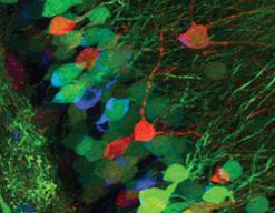
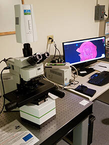
Zeiss LSM 800 confocal miscroscope with Airyscan
This microscope features classical confocal point scanning capability for optical sectioning and 3D reconstruction as well as sub-diffraction resolution capability through the Airyscan detector. Adjustable spectral filters and 4 lasers make this a suitable system for almost any dye in the visible range. An environmental chamber with temperature, humidity, and gas mixture control allows extended observation of live samples.

Rarecyte Orion for spatial biology (new!)
This multispectral spatial biology system uses narrow filtersets to image up to 20 unique fluorescently tagged biomarkers without cyclic staining. This system is great for visually validating Xenium or RNAseq targets at the protein level. It is optimized for Argofluor-labeled panels available from Rarecyte’s website.
Akoya Vectra 3 automated multispectral pathology imaging system
This system is an upright automated slide scanner for scanning of up to six slides with chromogenic (i.e., H&E, IHC) or fluorescence staining. The instrumentation is capable of multi-spectral imaging and spectral unmixing, which dramatically decreases background and allows for a high degree of multiplexing of a broad range of fluorophores in the same sample. Vectra, Phenochart, inForm, and HALO3 software is available for acquisition control and image analysis.
Cytation 5 automated imager with BioSpa8 incubator

This system houses up to 8 rectangular cell culture plates in a CO2 cell culture incubator and can image with brightfield, phase contrast, and up to four channels of florescence. Nitrogen is available to maintain hypoxic conditions for an additional cost. The system is ideal for performing high throughput long-term experiments on live samples without disrupting culture conditions.

Olympus IX83 / PRIMO micropatterning microfabrication
This microscope serves as a platform for a PRIMO bioengineering device. The PRIMO is capable of precise UV light-based patterning of molecules on any surface and microfabrication using hydrogels based over ranges from micron-scale features to centimeter-scale culture areas. An Andor Sona sCMOS camera allows alignment with sample features and monitoring of pattern progress.
Olympus IX73 widefield microscope

This is a versatile manual widefield inverted microscope for routine fluorescence and colored brightfield imaging. It is equipped with long working distance objectives for imaging thick plates/coverslips, optics for phase contrast, and polarized light microscopy, as well as a near-infrared (800nm) filter set.
Image analysis workstation
We offer an independent workstation for post-acquisition analysis featuring multiple image analysis packages licenses including HALO, InForm, and Gen5, as well as serving as a platform for open-source analysis workflows.