


Location
Third level of Borwell in the Animal Resource Center
(603) 646-2757
Dartmouth Cancer Center
One Medical Center Drive
Lebanon, NH 03756
Pricing information and sample submission
The mission of the Dartmouth Cancer Center Imaging and Irradiation Shared Resource is to:
- Assist investigators in radiation treatment planning and the delivery of ionizing irradiation to cells, rodents, large animals and spontaneous animal tumors
- Provide technologies for rodent and large-animal imaging used in pre-clinical research studies
Please acknowledge the Microscopy resource of the Dartmouth Cancer Center Irradiation, Imaging, Microscopy and Animal Cancer Models Shared Resource (RRID:SCR_025077) along with NCI Cancer Center Support Grant 5P30CA023108, in any publications or presentations.
Leadership

P. Jack Hoopes DVM, PhD
Co-Director, Imaging
P.Jack.Hoopes@Dartmouth.edu
(603) 650-5031
Research Interest: Experimental cancer therapeutics including radiation, chemotherapy, hyperthermia, photodynamic therapy and nanotechnology (iron oxide nanoparticle hyperthermia cancer treatment), animal pathology, animal models, experimental surgical technology.

David J Gladstone, ScD
Co-Director, Irradiation
David.J.Gladstone@dartmouth.edu
(603) 650-6442
Research interest: Imaging, radiation physics, radiation treatment planning

Diana J. Wallin, PhD
Research Scientist
Diana.J.Wallin@hitchcock.org
Research interest: Imaging, anesthesia, animal research techniques, neurobiology
Services
Small animal imaging
- Varian Direct- Drive 9.4T MRI Research Scanner. Primary Use: pre-clinical imaging studies
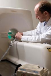
- Philips Achieva 3.0T MRI Clinical Scanner. Primary Use: pre-clinical imaging studies and MRI
- VisualSonics Vevo High-Frequency Ultrasound. Primary Use: high detail real-time ultrasound studies, cardiac output, and tumor volume estimates
- Xenogen VivoVision IVIS Bioluminescent and Fluorescent Imager. Primary Use: imaging bioluminescent and fluorescent reporters both in-vivo and in-vitro
- Philips MOSAIC micro-PET Scanner. Primary Use: radioisotope-based molecular imaging
- GE eXplore Locus In-Vivo micro-CT Scanner. Primary Use: highly detailed in-vivo imaging studies
- GE eXplore Locus Specimen micro-CT Scanner. Primary Use: highly detailed ex-vivo imaging studies
- GE Lightspeed CT Clinical Scanner. Primary Use: imaging studies and radiation therapy treatment planning
- OEC Cardiac 9800 Cine-Fluoroscope. Primary Use: real-time radiographic imaging
- FLIR SC325 Thermal Camera- Primary Use: real-time surface thermography
Large animal imaging
- Center for Surgical Innovation – Intraoperative MR, CT, and Bi-planar fluoroscopy
- GE Lightspeed CT Clinical Scanner. Primary Use: imaging studies and radiation therapy treatment planning
- Philips Achieva 3.0T MRI Clinical Scanner. Primary Use: pre-clinical imaging studies and MRI
- Philips 7500 Clinical Ultrasound. Primary Use: 3-D capable of cardiac imaging
- FLIR SC325 Thermal Camera- Primary Use: real-time surface thermography
Contact
For information, please contact:
- David Gladstone at David.J.Gladstone@dartmouth.edu or (603) 650-6600
- P. Jack Hoopes at P.Jack.Hoopes@dartmouth.edu or (603) 646-2757
- Diana J. Wallin at Diana.J.Wallin@hitchcock.org
Turnaround time and quality
Quality assurance-specific QA/QC procedure, for each of the irradiator services and instruments.
Irradiators operated in compliance with licensing requirements stipulated by the State of New Hampshire's Department of Health and Human Services Radiation Division, which regulates such source on behalf of the U.S. Nuclear Regulatory Commission (NRC). Dartmouth's Radiation Safety Officer, Thomas Diaz (603) 646-1794 or Thomas.J.Diaz@Dartmouth.edu